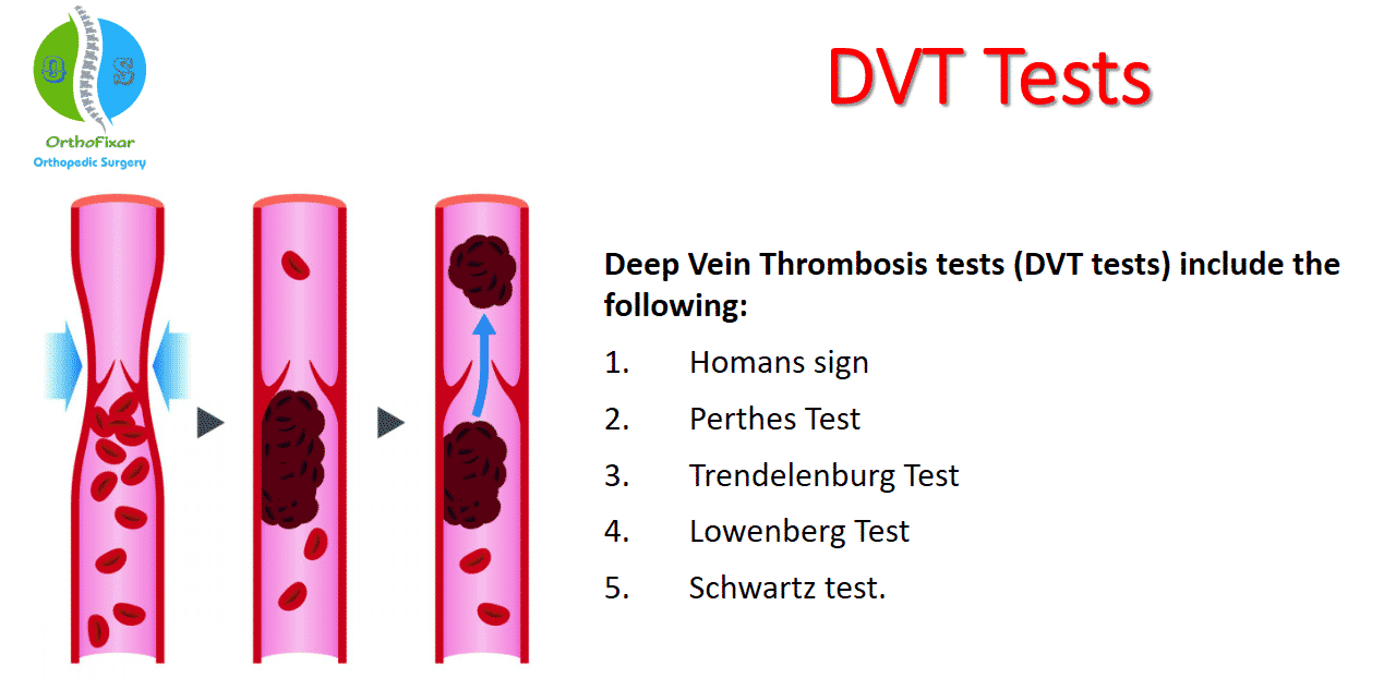
Deep Vein Thrombosis (DVT) is a serious medical condition that occurs when a blood clot forms in the deep veins of the body, most commonly in the legs. If left untreated, it can lead to life-threatening complications like pulmonary embolism, where the clot breaks loose and travels to the lungs. Understanding the diagnostic process for DVT, the tests involved, and what to expect during a medical evaluation is crucial for early detection and treatment.
In this comprehensive guide, we’ll explore the various steps involved in diagnosing DVT, the tests you might undergo, and what signs and symptoms to watch for to ensure early intervention.
Why Is Early Diagnosis Critical?
Deep Vein Thrombosis is the formation of a blood clot (thrombus) in the deep veins, typically in the legs but also potentially in other areas like the arms or pelvis. These clots restrict blood flow and can cause pain, swelling, and other complications. The condition becomes especially dangerous if the clot dislodges and travels to the lungs, causing a pulmonary embolism, which can be fatal.
Early diagnosis is critical because it can prevent the clot from growing larger or breaking off and migrating to the lungs. DVT diagnosis typically involves a combination of clinical evaluation, imaging tests, and sometimes blood work to confirm the presence of a clot.
Common Symptoms of DVT: When to Seek a Test
Recognizing the signs of DVT is essential for knowing when to seek a test. The most common symptoms include:
1. Pain and Tenderness in the Leg
One of the earliest signs of DVT in the leg is pain, often described as a cramping or aching sensation. The pain usually starts in the calf but can extend upward toward the thigh, especially if the clot is higher up in the leg. Deep vein thrombosis calf symptoms are often confused with muscle strains or cramps, but DVT pain tends to worsen over time rather than improving with rest or stretching.
2. Swelling in One Leg
Swelling is another classic symptom of DVT. It usually affects only one leg, although in rare cases, both legs may be involved. The swelling can be significant, making the leg feel tight and heavy. The swelling occurs because the blood clot blocks the flow of blood, causing it to pool in the leg.
3. Warmth and Redness
The skin over the affected area may become warm to the touch and red or discolored. This occurs as a result of the blocked blood flow, which leads to an increase in pressure within the veins.
4. Visible Veins
In some cases, the veins near the surface of the skin may become more visible or appear twisted. This is a result of the body trying to reroute blood around the blocked vein.
If you experience any combination of these symptoms, it’s important to seek medical attention immediately for a DVT diagnosis.
Diagnostic Tests for Deep Vein Thrombosis
Once you’ve consulted a healthcare provider, they will assess your symptoms and medical history before recommending specific tests to confirm whether you have DVT. Here are the most common tests used for deep vein thrombosis diagnosis:
1. Physical Examination and Medical History
Your doctor will start with a thorough physical examination, looking for visible signs of DVT such as swelling, redness, or warmth in the affected leg. They will also ask about your medical history, including any recent surgeries, long periods of immobility, or a family history of blood clots.
2. D-Dimer Test
The D-Dimer test is a blood test that measures the level of a substance released when a blood clot breaks down. Elevated D-dimer levels can indicate the presence of an abnormal blood clot, but this test is not definitive. If the D-dimer levels are normal, it is unlikely that you have DVT. However, elevated levels alone are not enough to diagnose DVT, as other conditions can also cause high D-dimer levels.
3. Duplex Ultrasound
A duplex ultrasound is the most commonly used test for diagnosing DVT. This non-invasive procedure uses sound waves to create images of the veins and detect the presence of a clot. During the test, a technician will use a handheld device (transducer) to scan the affected leg, looking for any blockages or abnormalities in the blood flow.
- How It Works: The ultrasound sends high-frequency sound waves into the leg, and these waves bounce back to the transducer to create an image of the blood vessels.
- What to Expect: The test is painless and typically takes about 30 minutes. You may feel some pressure as the technician moves the transducer over the leg, but it is generally a comfortable procedure.
4. Venography
In cases where the ultrasound results are unclear or the clot is located in a less accessible area, a venography may be recommended. This is a more invasive test that involves injecting a contrast dye into a vein, usually in the foot or ankle, and taking X-rays to visualize the veins.
- How It Works: The contrast dye allows the veins to be seen more clearly on the X-rays, helping to detect any blockages or clots.
- What to Expect: You may feel a warm sensation when the dye is injected, but the procedure is generally well-tolerated. Venography carries a small risk of allergic reaction to the dye and is less commonly used due to advances in ultrasound technology.
5. Magnetic Resonance Imaging (MRI) or Computed Tomography (CT) Scan
For cases where the clot is suspected in areas other than the legs, such as the pelvis or abdomen, an MRI or CT scan may be used. These imaging techniques provide detailed pictures of the blood vessels and can help identify clots in hard-to-reach areas.
- What to Expect: Both MRI and CT scans are non-invasive procedures that take detailed images of your blood vessels. These tests can take longer than ultrasounds, and in some cases, you may need an intravenous contrast dye for clearer images.
Understanding the Different Types of DVT
Deep Vein Thrombosis can occur in different parts of the body, and the symptoms and risks may vary depending on the location of the clot. Here’s an overview of the different types of DVT and their associated symptoms:
1. Calf DVT
The most common type of DVT occurs in the calf muscles. Deep vein thrombosis calf symptoms include localized pain, swelling, and tenderness. Calf DVT is often the result of prolonged immobility, such as during long flights or bed rest. While calf DVTs are usually smaller, they can still grow and cause serious complications if left untreated.
2. Thigh DVT
DVT can also occur in the thigh, where it may cause more severe pain that extends upward toward the groin. Deep vein thrombosis in thigh symptoms often include a dull ache, heaviness, and swelling in the upper leg. Thigh DVTs are generally considered more dangerous than calf DVTs because they are closer to the body’s larger veins, increasing the risk of the clot breaking off and traveling to the lungs.
3. Upper Extremity DVT
Although less common, DVT can also occur in the arms. Upper extremity DVT may cause symptoms such as swelling, pain, and discoloration in the arm, shoulder, or neck. This type of DVT is often associated with medical procedures like catheter placements or pacemaker insertions.
Risk Factors for DVT
Certain factors increase the likelihood of developing DVT. These include:
- Prolonged Immobility: Long periods of sitting, such as during travel or bed rest, can slow down blood flow and lead to clot formation.
- Surgery or Injury: Major surgeries, especially those involving the legs or hips, increase the risk of developing blood clots. Injuries to the blood vessels can also trigger clotting.
- Medical Conditions: Chronic conditions such as heart disease, cancer, and inflammatory bowel disease are known to increase the risk of DVT.
- Hormone Therapy: Hormone replacement therapy and birth control pills that contain estrogen are linked to a higher risk of blood clots.
- Obesity: Excess weight puts added pressure on the veins in the legs, increasing the risk of DVT.
- Genetics: A family history of DVT or blood clotting disorders can increase your risk of developing the condition.
Medical Treatment After a DVT Diagnosis
Once a DVT diagnosis has been confirmed, prompt treatment is essential to prevent the clot from growing or breaking off and traveling to the lungs. Here’s what you can expect after a diagnosis:
1. Anticoagulant Medications
The most common treatment for DVT is anticoagulant medication, also known as blood thinners. These medications, such as warfarin or heparin, help prevent the clot from growing and reduce the risk of new clots forming. However, they do not dissolve existing clots; the body will break down the clot naturally over time.
2. Thrombolytic Therapy
In more severe cases, where the clot is large or causing significant blockage, thrombolytic medications may be administered to dissolve the clot more quickly. This treatment carries a higher risk of bleeding and is typically reserved for life-threatening cases.
3. Compression Stockings
To help reduce swelling and prevent complications like post-thrombotic syndrome, compression stockings may be recommended. These stockings improve blood flow in the legs and help prevent further clots from forming.
4. Surgical Intervention
In rare cases, surgery may be required to remove a clot, especially if it is causing significant blockages in critical veins. This procedure is known as thrombectomy.
Conclusion: The Importance of DVT Testing for Early Detection
Recognizing the symptoms of DVT early and undergoing the appropriate deep vein thrombosis test is key to preventing complications and ensuring effective treatment. If you experience any of the warning signs, such as deep vein thrombosis pain or swelling, consult your healthcare provider promptly for a DVT diagnosis. Tests like ultrasound and D-dimer can confirm the presence of a clot and guide your treatment plan.
